Topic 1: Photon-Counting CT:
Improving Coronary Imaging in Patients with Heavy Calcification and Coronary
Stents

Figure 1. Photon-counting CT reduces image artifacts caused
by coronary calcification and metallic stents, allowing clearer visualization
of the coronary lumen and improved detection of in-stent restenosis. (Image
courtesy of Siemens Healthineers.)
Does chest tightness or chest pain always mean a blocked coronary artery?
Many patients undergo coronary computed tomography angiography (CCTA) to
evaluate their coronary arteries. However, when there is extensive coronary
artery calcification or a previously implanted coronary stent, conventional CT
scanners may be affected by calcium and metal-related artifacts, commonly known
as the "blooming artifact."
These artifacts can make the arterial lumen appear narrower than it actually
is, making it difficult to determine whether a true blockage exists. As a
result, some patients still need to undergo invasive coronary angiography for a
definitive diagnosis.
The latest photon-counting CT
(PCCT) offers significantly higher spatial resolution and effectively
reduces image artifacts caused by coronary calcification and metallic stents.
This enables physicians to visualize the coronary artery lumen more clearly.
Even in patients with high coronary calcium scores or previously implanted
stents, PCCT provides a more accurate assessment of the severity of coronary
artery narrowing and helps detect in-stent
restenosis. Studies have also shown that this technology improves
diagnostic accuracy, reduces unnecessary invasive coronary angiography, and
allows patients who require treatment to receive appropriate care more
promptly.
If you experience chest
discomfort or chest pain, or have risk factors such as hypertension, diabetes,
hyperlipidemia, smoking, or a history of coronary stent implantation, consult
your physician about whether coronary CT angiography is appropriate for you. At
Far Eastern Memorial Hospital,
the latest photon-counting CT technology provides high-resolution, non-invasive
cardiovascular imaging, enabling clearer visualization of the coronary arteries
and more accurate diagnosis.
Topic:2 Spectral CT: Revealing Small Renal Tumors
with Greater Confidence
Figure
2.
The small renal tumor is difficult to appreciate on a conventional CT image
(left). Spectral CT low-energy images (center) and iodine maps (right) provide
clearer visualization of the lesion and its blood supply. (Image courtesy of Siemens
Healthineers.)
Some
renal tumors are very small or have an attenuation similar to that of normal
kidney tissue, making them difficult to detect on conventional CT scans.
The
latest Spectral CT
technology goes beyond conventional CT imaging by analyzing X-rays at different
energy levels to generate low-energy
images and iodine maps. These
advanced images allow radiologists to determine whether a lesion truly enhances
after contrast administration, providing valuable information about its blood
supply. This helps differentiate tumors from cysts and other benign lesions
with greater confidence.
For
patients, the greatest benefit is improved diagnostic confidence. More accurate
imaging can reduce the need for repeat examinations, minimize delays in
diagnosis, and help physicians develop appropriate treatment plans at an
earlier stage.
Far
Eastern Memorial Hospital has introduced the latest Spectral CT technology to
improve the detection of small lesions in the kidneys, liver, pancreas, and
other abdominal organs. By providing more precise diagnostic information, this
advanced imaging technology supports earlier diagnosis and more personalized
patient care.
High-Resolution,
Low-Dose Chest CT with Photon-Counting CT
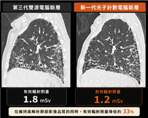
Figure
3.
High-resolution, low-dose photon-counting CT provides clearer visualization of
pulmonary fibrosis while reducing radiation exposure. (Image courtesy of Siemens
Healthineers.)
Many
people worry about radiation exposure from CT examinations and may delay evaluation
of lung diseases because of these concerns. Fortunately, advances in CT
technology now make it possible to achieve both higher image quality and lower radiation doses.
For
conditions such as pulmonary
fibrosis, emphysema, and lung
cancer screening, the latest photon-counting
CT (PCCT) offers ultra-high-resolution images that reveal
fine lung structures in greater detail. This allows radiologists to detect
subtle abnormalities more easily while reducing radiation exposure by
approximately 30%.
For patients who require long-term follow-up of lung diseases, PCCT not only
improves diagnostic accuracy but also minimizes cumulative radiation exposure.
If
you have a persistent cough, shortness of breath, a history of smoking, or a
family history of lung cancer, talk with your physician about whether a chest
CT examination is appropriate for you.
At Far Eastern Memorial Hospital, the latest high-resolution, low-dose
photon-counting CT technology provides more accurate diagnosis of lung diseases
while maintaining excellent image quality and reducing radiation exposure,
helping patients receive timely and appropriate care.