Specialties: Da Vinci-assisted minimally invasive lung
and mediastinal surgery, 3D thoracoscopic minimally invasive surgery, lung
diseases, mediastinal diseases, pectus excavatum, chest wall tumors, rib
fracture reduction surgery, trauma, adjuvant cancer treatment
What Is Pneumothorax?
Pneumothorax
is a condition caused by rupture of the alveoli, allowing air to accumulate in
the pleural space. This trapped air prevents the lung from fully expanding and
may compress the lung, leading to partial or complete collapse.
When the lung is significantly compressed, patients
may suddenly experience chest pain, chest tightness, shortness of breath,
and a sensation of being unable to inhale sufficient air,
potentially resulting in hypoxia. Some patients may also develop cough, shoulder
pain, neck discomfort, or upper back soreness.
If a large volume of air accumulates rapidly, it may
cause mediastinal
shift, impaired venous return to the heart, hypotension, and shock.
This life-threatening condition is known as tension pneumothorax
and requires immediate emergency treatment.
Types and Causes of
Pneumothorax
In addition to traumatic pneumothorax caused by blunt
or penetrating chest injury, primary spontaneous pneumothorax
commonly occurs in young, tall, slender, otherwise healthy males, often due
to rupture of apical lung blebs or bullae.
Although the exact mechanism is not fully understood,
forceful
coughing, sneezing while covering the nose and mouth, and cigarette smoking
significantly increase the risk of bleb formation and rupture. Smoking cessation is
the most important preventive measure.
Secondary spontaneous pneumothorax is
more frequently seen in older patients or those with underlying lung diseases
such as emphysema, chronic obstructive pulmonary disease (COPD), pulmonary
tuberculosis, pneumoconiosis, or cystic fibrosis.
Clinical Symptoms and Warning
Signs
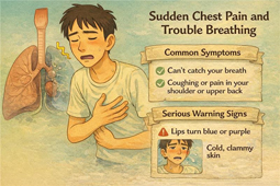
Common symptoms of pneumothorax include:
·
Sudden onset chest pain or chest
tightness
·
Shortness of breath or difficulty breathing
·
Cough,
shoulder pain, or back pain
·
In severe cases: cyanosis, cold
sweats, tachycardia, or hypotension
Patients presenting with severe chest
tightness, low blood pressure, or altered consciousness should
seek immediate medical attention due to the risk of tension pneumothorax.
Treatment Goals of
Pneumothorax
The primary goals of treatment are to:
1.
Relieve symptoms
2.
Promote lung healing
3.
Prevent recurrence
Most mild pneumothoraces may resolve spontaneously.
However, patients with severe symptoms, lung collapse greater than 20%, or
persistent air leakage require chest tube drainage and
possibly surgical intervention.
Management of First-Episode
Unilateral Primary Spontaneous Pneumothorax
·
Lung collapse < 20%
with mild symptoms:
→ Outpatient observation with instructions to return if symptoms worsen
·
Lung collapse > 20%
or dyspnea present:
→ Placement of a pigtail catheter for drainage and monitoring
·
Persistent air leak or failure
of lung re-expansion after 48 hours:
→ Consider video-assisted thoracoscopic surgery (VATS)
Indications for Primary
Surgical Treatment
Immediate or early surgery is recommended in the following
situations:
·
Life-threatening conditions:
pneumothorax with hemothorax, bilateral pneumothorax
·
Second ipsilateral recurrence
or contralateral pneumothorax
·
Persistent air leak after chest
tube insertion
·
High-risk occupations (e.g.,
pilots, flight attendants, divers)
·
Limited access to medical care
or remote residence
VATS and Pleurodesis
Video-assisted thoracoscopic surgery (VATS) is currently the most effective method for reducing recurrence. The
procedure involves:
·
Identification and resection or
ligation of lung bullae (bullectomy)
·
Pleurodesis, which promotes adhesion between the lung and chest wall
Pleurodesis techniques include:
1.
Mechanical pleurodesis – pleural abrasion or partial pleurectomy
2.
Chemical pleurodesis – instillation of agents such as talc, minocycline, OK-432, or
hypertonic glucose
3.
Artificial pleural mesh coverage (commonly used in our institution), which thickens the pleural
surface and reduces recurrence with fewer side effects such as fever or pain
Recent studies demonstrate that pleural mesh coverage
for postoperative air leak is safe, effective, shortens hospital stay, and lowers
recurrence rates.
Postoperative Care and
Recurrence Prevention
To reduce the risk of recurrence, patients should:
·
Avoid strenuous exercise and
forceful coughing for 3 months
·
Attend scheduled follow-up
visits with chest X-rays at 2 weeks, 1 month, 3 months, and 6 months
·
Completely stop smoking
Conclusion
Primary spontaneous pneumothorax is an acute but
highly treatable thoracic condition. With appropriate management, most patients
can fully
recover and return to normal daily activities. VATS combined with
pleurodesis remains the most effective strategy for preventing
recurrence. Adherence to postoperative care, smoking cessation, and regular
follow-up significantly improve quality of life and long-term outcomes.
References:
O
How CH, Tsai TM, Kuo SW, Huang PM, Hsu HH, Lee
JM, Chen JS, Lai HS. Chemical pleurodesis for prolonged postoperative air leak
in primary spontaneous pneumothorax. J Formos Med Assoc. 2014
May;113(5):284-90.
O
Hsu HH, Liu YH, Chen HY, Chen PH, Chen KC, Hsieh
MJ, Lin MW, Kuo SW, Huang PM, Chao YK, Wu CF, Wu CY, Chiu CH, Chen WH, Wen CT,
Liu CY, Wu YC, Chen JS. Vicryl Mesh Coverage Reduced Recurrence After
Bullectomy for Primary Spontaneous Pneumothorax. Ann Thorac Surg. 2021
Nov;112(5):1609-1615.
O
MacDuff A, Arnold A, Harvey J; BTS Pleural
Disease Guideline Group. Management of spontaneous pneumothorax: British
Thoracic Society Pleural Disease Guideline 2010. Thorax. 2010 Aug;65 Suppl
2:ii18-31. doi: 10.1136/thx.2010.136986. PMID: 20696690.
O
Cheng HS, Wong C, Chiu PH, Tong CW, Miu PF.
Management of spontaneous pneumothorax: a mini-review on its latest evidence. J
Thorac Dis. 2024 Jul 30;16(7):4756-4763.