
Specialties: Coronary artery disease and myocardial infarction, cardiac catheterization, balloon angioplasty and stent placement, peripheral vascular disease, and heart failure.
In
clinical practice, we often encounter a rapidly developing, unpredictable, and
potentially fatal condition: Deep Vein Thrombosis (DVT) and Pulmonary Embolism
(PE). These two conditions fall under the category of venous thromboembolism.
Though they may appear harmless on the surface, they often strike without
warning, catching both patients and their families off guard, and in some
cases, can be fatal. From a physician’s perspective, these diseases require
prompt identification and treatment, and more importantly, should be well
understood by the general public to reduce the risk of delayed care.
As the name
suggests, deep vein thrombosis refers to the abnormal formation of blood clots
within the deep veins of the body, most commonly in the veins of the calf or
thigh. When these clots dislodge, they can travel through the bloodstream to
the lungs and block a pulmonary artery, resulting in a pulmonary embolism. This
is a life-threatening emergency that can lead to respiratory failure, low blood
pressure, or sudden death.
The
formation of blood clots is typically associated with three major mechanisms.
The first is blood flow stasis, which can occur due to prolonged sitting, bed
rest, or reduced mobility after surgery. The second is damage to the vessel
wall, often due to trauma, surgical procedures, or infections. The third is
hypercoagulability of the blood, which may be related to cancer, pregnancy,
hereditary conditions, or hormone therapies. These factors may exist
independently or in combination to contribute to thrombus formation.
Many
patients do not notice anything unusual at first, only seeking medical
attention when their leg becomes swollen, painful, warm, or tender. However,
the more serious manifestations often arise from a pulmonary embolism. Some
individuals may suddenly experience shortness of breath, chest tightness or
pain, rapid heartbeat, or dizziness. These symptoms are frequently mistaken for
heart disease, asthma, or anxiety, leading to delays in seeking treatment. Even
more concerning, some patients’ first symptom is sudden death, highlighting the
critical importance of early recognition and prevention.
For
diagnosis, physicians rely on the patient’s medical history, symptoms, and
clinical risk assessments to determine the appropriate tests. Initial blood
tests such as D-dimer can help rule out low-risk cases, while ultrasound can
directly visualize blood flow in the leg veins. If PE is suspected, Computed
Tomography Pulmonary Angiography (CTPA) is the most commonly used and accurate
imaging modality, clearly showing the location and severity of the clot. These
tests are essential for making a prompt diagnosis and evaluating treatment
options.
Once
a diagnosis is confirmed, the first line of treatment usually involves
anticoagulant medications. These drugs effectively prevent the clot from
growing and stop new clots from forming. Traditionally, low-molecular-weight
heparin and warfarin have been used. In recent years, however, more patients
have been treated with novel oral anticoagulants (NOACs) such as apixaban or
rivaroxaban, which offer convenience and do not require frequent blood
monitoring. For patients with large clots or unstable vital signs, doctors may
consider hospitalization and thrombolytic therapy to dissolve the clots,
although these medications carry a higher risk of bleeding and must be used
with caution.
In
particularly severe cases—such as when a clot blocks a major pulmonary artery,
causing severe hypoxia, hypotension, or even shock—conventional medical
treatment may not work quickly enough. In such situations, Catheter-Directed
Thrombectomy becomes a critical interventional option. This minimally invasive
technique, which has rapidly advanced in interventional radiology and
cardiovascular medicine at our hospital in recent years, offers the benefits of
safety, speed, and reduced invasiveness.
The principle behind catheter-directed
thrombectomy is to insert a thin catheter through a blood vessel in the groin
or neck, navigate it through the vascular system to the site of the pulmonary
embolism, and use negative pressure or mechanical devices to extract the clot.
Common devices include the Penumbra Indigo and AngioJet systems, both of which
can remove blockages quickly, restoring pulmonary blood flow and improving
oxygenation and blood pressure. Most importantly, this procedure typically does
not require high doses of thrombolytic drugs, significantly reducing the risk
of intracranial or other major bleeding. We have already successfully performed
catheter-directed thrombectomy on several patients, saving lives—particularly
in high-risk individuals who cannot receive thrombolytics, such as those with
prior strokes, active bleeding, or advanced age. For these patients, catheter
intervention provides a vital, life-saving alternative.
Despite
advancements in treatment technology, prevention remains even more crucial. If
you often sit for long periods, take long-haul flights, or have recently
undergone surgery or hospitalization, it is essential to move your limbs, wear
compression stockings, stay hydrated, and consult your doctor about the need
for preventive medication. Individuals with a family history of thrombosis,
cancer patients, or pregnant women should also remain vigilant—prevention is
always better than cure.
In
the world of medicine, DVT and PE may not be the most talked-about conditions,
but they are among the most dangerous threats we face. With proper awareness,
effective treatment, and proactive care for high-risk populations, we can
greatly reduce complications and mortality. The smooth flow of every blood
vessel is the foundation of life stability, and every alertness to thrombosis
is the first step in safeguarding health.
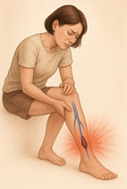 Deep Vein Thrombosis (DVT) schematic diagram
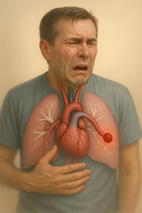 Pulmonary Embolism (PE) schematic diagram
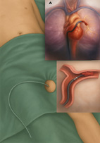 Diagram illustrating the catheter suction proc
 edure
edure